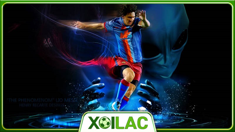
Xem bóng đá Xoilac chất lượng 4K, không quảng cáo
Truy cập ngay hôm nay để xem bóng đá Xoilac với chất lượng cao. Trải nghiệm những trận đấu đỉnh cao đến từ nhiều giải đấu hàng đầu quốc tế.
Ngày 26/04/2024
Trang xem bóng đá Xoilac full HD không bị đứng hình, phát sóng trực tiếp toàn bộ giải đấu lớn nhỏ trong nước và quốc tế. Xem trực tiếp bóng đá hoàn toàn miễn phí trên trang Xôi Lạc. Một trong những nền tảng chuyên biệt cung cấp link theo dõi trận đấu chất lượng và ổn định nhất hiện nay. Cùng tìm hiểu những tính năng nổi bật của kênh trong bài viết dưới đây.
Link truy cập dành cho IOS/Android
Đối với những người muốn truy cập xem bóng đá Xoilac trên thiết bị IOS hoặc Android, các liên kết chính thức dưới đây đảm bảo một cách nhanh chóng và thuận tiện để tham gia vào thế giới bóng đá hấp dẫn:
- Xoilac 1
- Xoilac 2
- Xoilac 3
- Xoilac 4
Để làm cho trải nghiệm trên nền tảng trở nên hoàn hảo hơn, đội ngũ Xoilac cam kết giải quyết những hạn chế và cung cấp các cập nhật đều đặn. Người dùng có thể mong đợi một trải nghiệm mượt mà hơn sau mỗi lần truy cập. Đối với những ai yêu thích những trận đấu đỉnh cao của nhiều câu lạc bộ lớn. Bạn đều có thể dễ dàng trải nghiệm
Đánh giá ưu điểm trang xem bóng đá Xoilac
Trang web mang đến cho người dùng của mình nhiều tính năng tuyệt vời, bạn sẽ không thể nào thất vọng khi tận hưởng những trận đấu mà mình yêu thích tại đây.
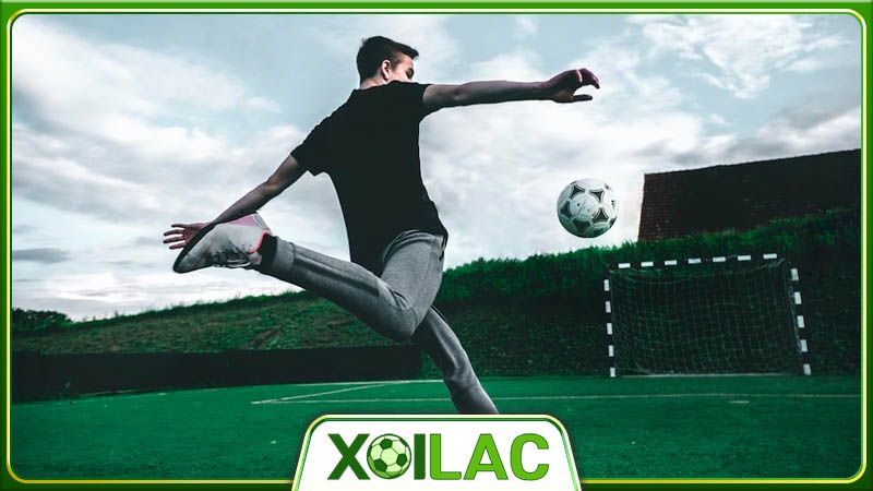
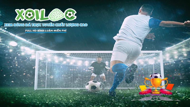
Xem bóng đá Xoilac full HD
Khám phá thế giới bóng đá với chất lượng hình ảnh tuyệt vời, trang web tự tin cam kết mang đến trải nghiệm xem trực tiếp bóng đá đỉnh cao qua độ phân giải Full HD. Tại đây, mỗi trận đấu được tái hiện một cách rõ ràng và chân thực, đưa người xem đến gần với sân cỏ hơn bao giờ hết.
Xôi Lạc TV không chỉ đặt ra mục tiêu cung cấp chất lượng hình ảnh Full HD, mà còn là biểu tượng của sự chăm sóc tận tình dành cho người hâm mộ và là sự khẳng định vững chắc về đam mê và tình cảm của kênh này đối với môn thể thao vua. Việc này không chỉ là hứa hẹn cho một trải nghiệm xem bóng đá, mà là một lời cam kết từ trái tim của kênh.
Bình luận công khai và an toàn
Với việc tích hợp tính năng bình luận trực tiếp trong trận đấu, xem bóng đá Xoilac tạo ra một không gian cho người xem không chỉ để thưởng thức trận đấu mà còn để tự do bày tỏ quan điểm của mình. Điều này không chỉ là một tính năng, mà là cơ hội cho mỗi người hâm mộ để giao tiếp, chia sẻ cảm xúc và tận hưởng không khí đam mê của bóng đá.
Quan trọng hơn trang web cam kết đảm bảo tính an toàn và không gian bình luận không xung đột. Với sự quản lý chặt chẽ, mọi ý kiến được đưa ra một cách văn minh và tôn trọng. Điều này giúp xây dựng cộng đồng xem bóng đá tích cực, nơi mà mọi người có thể chia sẻ và kết nối qua đam mê chung của họ mà không lo lắng về giao tiếp tiêu cực.
Công nghệ trực tiếp hiện đại
Một trong những điểm mạnh của trang xem bóng đá Xoi Lac là khả năng kết hợp và cung cấp công nghệ phát sóng trực tiếp bóng đá đỉnh cao. Tạo nên một trải nghiệm độc đáo và tuyệt vời cho người hâm mộ. Kênh cam kết mang đến sự suôn sẻ và thú vị cho việc xem trực tiếp bóng đá trên trang. Đồng thời giảm thiểu tối đa tình trạng giật lag và đảm bảo chất lượng phát sóng mượt mà nhất cho người xem.
Xoilac TV không chỉ đơn thuần là một nơi xem trực tiếp trận đấu, mà còn là không gian của sự hiện đại và tiện ích. Đội ngũ kỹ thuật chúng tôi luôn làm việc chăm chỉ để nâng cao công nghệ phát sóng, giúp việc trải nghiệm xem bóng đá trực tiếp trở nên hoàn hảo hơn.
Chính sách bảo mật an toàn
Khi bước chân vào thế giới của trang xem bóng đá Xoilac, bạn hoàn toàn yên tâm về thông tin cá nhân của mình. Chính sách bảo mật an toàn của chúng tôi không chỉ là sự cam kết, mà là một bảo vệ vững chắc cho sự riêng tư của bạn. Tại đây, chúng tôi sử dụng những biện pháp bảo mật tiên tiến để mọi giao dịch diễn ra an toàn và bảo mật.
Đội ngũ CSKH chuyên nghiệp
Nền tảng này không chỉ là nơi bạn xem bóng đá, mà còn là một gia đình chân thành. Đội ngũ chăm sóc khách hàng của chúng tôi không chỉ là những nhân viên, mà là những người bạn đồng hành. Chúng tôi luôn sẵn sàng lắng nghe và giải quyết mọi vấn đề của bạn với tận tâm và tận tình, vì đối với chúng tôi, mỗi khách hàng là một phần quan trọng của cộng đồng Xoilac.
Giao diện dễ sử dụng
Khám phá thế giới bóng đá với giao diện dễ sử dụng tại đây. Trang web xem bóng đá Xoilac được thiết kế để đặt trải nghiệm của bạn lên hàng đầu. Giao diện tinh tế và trực quan giúp bạn thoải mái thưởng thức từng khoảnh khắc của trận đấu. Tại đây, chúng tôi không chỉ mang đến tính năng vượt trội, mà còn đảm bảo mỗi người xem đều có trải nghiệm mượt mà và không gặp khó khăn trong việc tương tác.
Kênh Xoialac phát sóng những giải đấu nào?
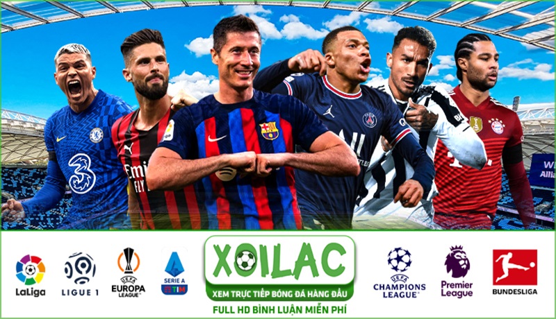
Cùng tìm hiểu liệu bạn có thể xem bóng đá Xoilac với những trận đấu hấp dẫn nào tại đây:
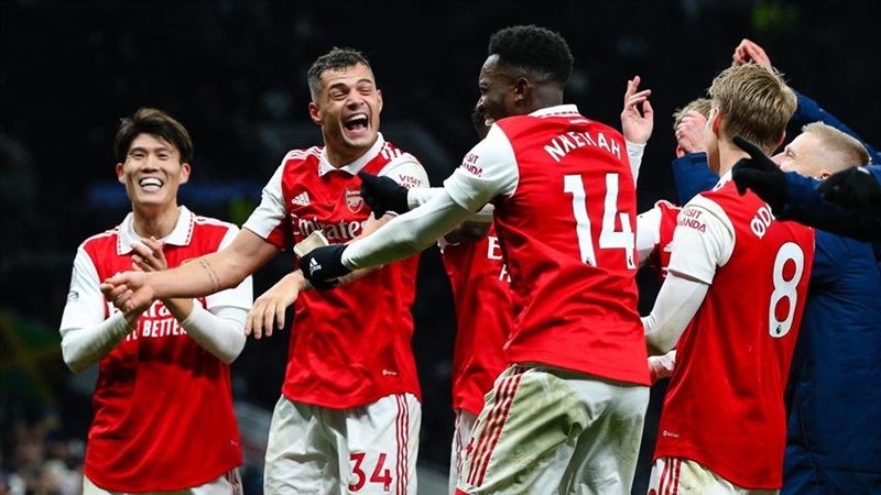
- Premier League: Xem những trận đấu căng thẳng và hấp dẫn nhất từ giải đấu hàng đầu nước Anh. Với những bàn thắng đẹp mắt sẽ làm bạn phấn khích và hài lòng.
- Euro – Bóng Đá Châu Âu: Với Euro, bóng đá châu Âu đưa bạn đến với những trận đấu không thể dự đoán và kịch tính. Xoilac là điểm đến đáng tin cậy để bạn không bỏ lỡ bất kỳ khoảnh khắc nào của giải đấu này.
- UEFA Champions League: Chinh phục đỉnh cao bóng đá châu Âu đây không chỉ là nơi phát sóng, mà còn là ngôi nhà của những trận đấu căng thẳng và những bàn thắng ấn tượng.
- World Cup: Khi World Cup diễn ra, trang web xem bóng đá Xoi Lac đã trở thành đối tác lý tưởng cho những người hâm mộ. Hãy thưởng thức những cảm xúc và niềm vui từ những trận đấu quốc tế tại đây.
- Bundesliga – Đức: Với Bundesliga mang đến không khí sôi động và tài năng của bóng đá Đức. Đội bóng mạnh mẽ và những pha bóng độc đáo sẽ làm cho mỗi trận đấu trở nên đáng nhớ.
- V-League: Trang web không quên đến với người hâm mộ Việt Nam với V-League. Những trận cầu đầy khí thế và lòng đam mê của bóng đá Việt Nam sẽ được phát sóng một cách chất lượng, làm hài lòng người xem.
Xoilac còn có tên gọi nào khác?
Xoilac, một kênh phát sóng trực tiếp bóng đá nổi tiếng, còn được biết đến với một số tên gọi khác như Xôi Lạc TV, Xôi Lạc 90 phút và 7m trực tiếp bóng đá. Những tên gọi này đã trở thành cái tên quen thuộc, gắn liền với sự đa dạng và phong phú của nội dung mà trang web mang đến cho người hâm mộ bóng đá.
Bên cạnh đó kênh xem bóng đá Xoilac cũng là nơi cung cấp những trận đấu hấp dẫn mà còn là nguồn cảm hứng, niềm đam mê vô tận đối với môn thể thao vua này. Không chỉ đơn thuần là một kênh truyền hình, đây cũng là người bạn đồng hành, đồng đội trung thành của người yêu bóng đá. Luôn sẵn sàng mang đến những trải nghiệm tuyệt vời và khám phá mới mẻ trong thế giới bóng đá.
Xem bóng đá Xoilac nên lưu ý gì?
Khi bạn quyết định xem bóng đá trên kênh Xoilac, hãy lưu ý những điều sau đây để có được nhiều trải nghiệm tuyệt vời và an toàn hơn. Cụ thể như:
- Trang web không có trách nhiệm về những nội dung được đăng tải từ những kênh khác.
- Nếu như bạn phát hiện có video vi phạm bản quyền, bạn có thể gửi yêu cầu đến hệ thống trang web đã được lưu trữ. Hoặc phát trực tiếp đến những chương trình đó, để họ chịu trách nhiệm.
- Kiểm tra kết nối internet để có được trải nghiệm tuyệt vời hơn.
- Bạn không cần đăng ký tài khoản khi xem bóng đá Xoilac.
- Người xem nên kiểm tra lịch thi đấu trước khi theo dõi trực tiếp trên kênh.
Một số câu hỏi thường gặp khi xem bóng đá Xoilac
Khi trải nghiệm trang web có rất nhiều vấn đề được nhiều người dùng quan tâm. Tất cả sẽ được chúng tôi giải đáp chi tiết trong nội dung dưới đây:
Xem bóng đá Xoilac có uy tín hay không?
Xoilac là một kênh phát sóng bóng đá trực tiếp có uy tín trong cộng đồng yêu bóng đá. Với chất lượng phát sóng cao và sự đa dạng trong nội dung, Xoilac đã giành được lòng tin của hàng triệu người hâm mộ. Chưa kể, kênh còn là trang có giấy phép hoạt động hợp pháp. Cho nên, bạn có thể hoàn toàn yên tâm khi trải nghiệm tại đây.
Có xuất hiện quảng cáo khi xem trực tiếp tại Xôi Lạc?
Không, Xoilac cam kết mang đến trải nghiệm xem bóng đá trực tuyến mượt mà và không gián đoạn. Kênh không xuất hiện quảng cáo làm phiền trước, trong, hoặc sau trận đấu, để bạn có thể tận hưởng mỗi khoảnh khắc của trận cầu mà không bị gián đoạn.
Tôi có mất phí khi trải nghiệm tại kênh không?
Không, trang xem bóng đá Xoilac cung cấp dịch vụ xem bóng đá trực tuyến hoàn toàn miễn phí. Bạn không cần trả bất kỳ chi phí nào để thưởng thức những trận đấu hấp dẫn và chất lượng tại đây.
Xoi Lac có link dự phòng không?
Để đảm bảo rằng người xem luôn có cơ hội truy cập, Xoilac thường cung cấp link dự phòng. Những link này sẽ giúp bạn tiếp cận nhanh chóng và đảm bảo rằng bạn không bỏ lỡ bất kỳ trận đấu quan trọng nào.
Xem bóng đá Xoilac ngoài việc giúp bạn đến gần hơn với những đội tuyển mình yêu thích. Bên cạnh đó, đây cũng là cách giải trí để bạn và gia đình có thể đoàn tụ sau những giờ làm việc hăng say. Hãy nhanh tay truy cập và tận hưởng những giải đấu mà mình yêu thích tại đây.